Published in Cell (2020), the study by Hou et al. delivers one of the most comprehensive looks at how SARS-CoV-2 infiltrates the human respiratory tract. By leveraging a sophisticated reverse genetics system and a suite of engineered reporter viruses, the researchers mapped the virus’s infectivity from the nose to the lungs—revealing an infection gradient that could hold the key to understanding COVID-19’s spread and severity.
Understanding the Virus: A New Reverse Genetics System
To study SARS-CoV-2 in unprecedented detail, the researchers constructed a full-length infectious cDNA clone of the virus, along with reporter viruses tagged with GFP and nanoluciferase. These allowed real-time visualization of infection and high-throughput testing of neutralizing antibodies from recovered patients and vaccines.
This toolkit enabled them to examine two key questions:
- Where does SARS-CoV-2 infect the respiratory tract most efficiently?
- How does antibody neutralization vary between SARS-CoV-2 and other coronaviruses like SARS-CoV and MERS?
Key Findings
1. A Steep Infection Gradient: Nose > Lungs
- Using RNA in situ hybridization and qPCR, the researchers showed that ACE2, the key receptor for SARS-CoV-2, is most highly expressed in the nasal epithelium and gradually decreases toward the alveoli.
- This correlated directly with infectivity: nasal epithelial cells supported the highest levels of viral replication, while alveolar cells showed the least.
2. Ciliated Airway Cells and AT2 Cells Are Primary Targets
- Within the respiratory tract, ciliated cells in the upper airway and type II alveolar cells (AT2) in the lungs were the main infection targets.
- Secretory and goblet cells, while present in infected areas, were not the primary sites of viral replication.
3. Cross-Neutralization Between Coronaviruses Is Limited
- Sera from SARS-CoV-1 (2003) and SARS-CoV-2 survivors showed limited cross-neutralization. Most SARS-specific monoclonal antibodies failed to neutralize SARS-CoV-2 effectively.
- This suggests that vaccines and therapeutics must be strain-specific, and that prior coronavirus infections may offer little protective immunity against SARS-CoV-2.
Why This Gradient Matters
The findings suggest that COVID-19 begins in the nose—where ACE2 expression and viral replication are highest—and may then seed the lungs through aspiration, especially during sleep or in high-risk populations like the elderly and obese.
This helps explain:
- Early nasal viral shedding, even before symptom onset
- Patchy, peripheral lung damage seen in autopsies and CT scans
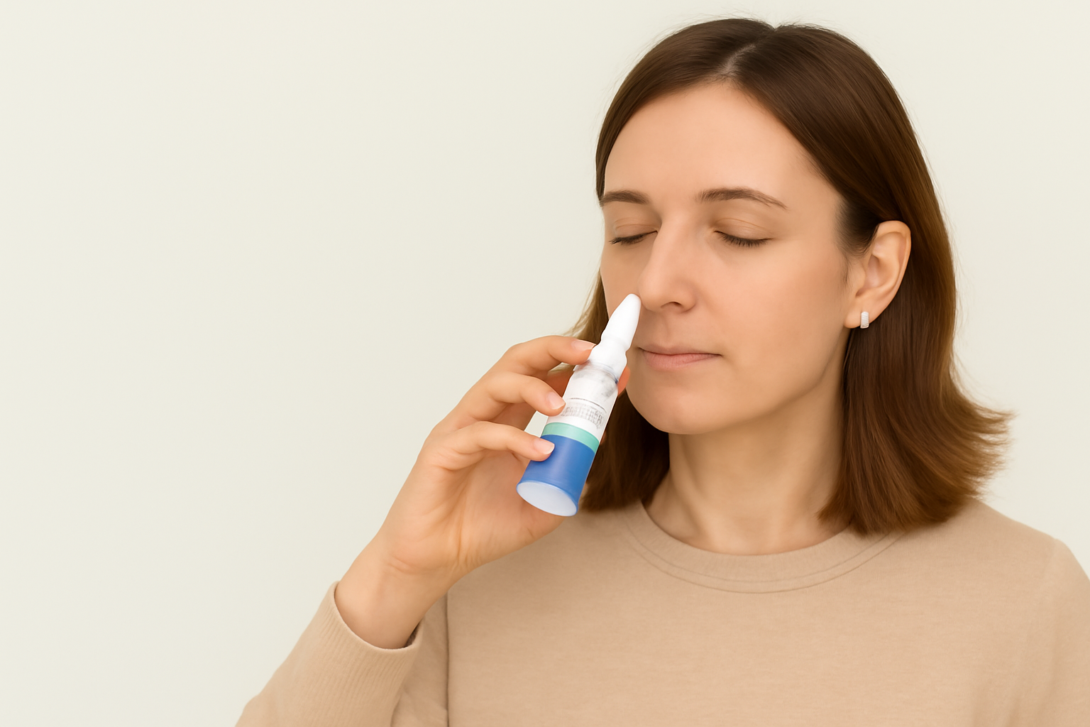
- Why nasal interventions (like topical antivirals or antiseptics) might be effective early in infection
The Role of Pre-existing Conditions
In patients with cystic fibrosis (CF) or other chronic lung diseases, ACE2 and TMPRSS2 were upregulated, possibly increasing vulnerability to SARS-CoV-2. However, the relationship is complex: ACE2 may also have protective anti-inflammatory effects in some contexts.
Cytokines like IL-1β and interferon-β upregulated ACE2 expression in vitro, while IL-13 (associated with asthma) suppressed it—further linking inflammation and susceptibility.
Implications for COVID-19 Prevention and Treatment
This study opens several promising avenues:
- Nasal-targeted therapeutics could block initial infection or reduce viral load early, especially in asymptomatic carriers.
- ACE2 expression profiles might help identify high-risk individuals or predict disease severity.
- Reverse genetics tools can streamline vaccine testing, neutralization assays, and drug development.
Conclusion
The nasal cavity is not just the virus's front door—it’s the control center for how COVID-19 unfolds in the body. Hou et al.’s work reveals a detailed map of SARS-CoV-2’s preferred terrain, highlighting the upper respiratory tract as the critical site for early infection and viral shedding.
Future efforts to control transmission, especially in community settings, may hinge on protecting the nose—with everything from masks and air filtration to nasal sprays and targeted antivirals.

.png)